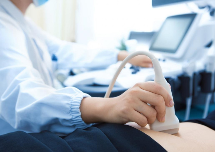
Medical diagnostic imaging forms the bedrock of accurate healthcare delivery, ensuring clinical interventions are guided by absolute precision rather than diagnostic speculation. At Ashwa Hospital, we provide state-of-the-art ultrasound scanning in Vijayawada, combining high-frequency sound wave technology with clinical expertise to view the internal architectures of the human body safely. As a leading specialized destination among gynecology hospitals in Vijayawada, our diagnostic infrastructure is optimized to map internal organs, monitor fetal developments, and evaluate complex vascular frameworks in real time without ionizing radiation exposure.
Related consultations: Patients in Vijayawada often also explore General Physician Consultations,Women’s Health Checkups,Nutrition Guidance During Pregnancy, Laparoscopic Surgeries based on symptoms and treatment goals.
Our advanced clinical ultrasound facility functions under the direct leadership of Dr. M. Madhavi, a highly regarded best gynecologist in Vijayawada, Consultant Obstetrician, Fertility Specialist, and Laparoscopic Surgeon. With extensive academic and practical experience, including her previous tenure as an Ex-Consultant at NRI Medical College, Dr. Madhavi utilizes clinical sonography as an interactive modality to resolve deep-seated women's health conditions and complex pregnancy paths.
Whether you require a baseline pelvic check, a sophisticated fetal anomaly clearance, an investigation into mysterious pelvic pains, or a transvaginal tracking matrix for an upcoming fertility cycle, our facility delivers unparalleled diagnostic clarity. Located accessibly on Dasarivari Street, Prakasam Road, Ashwa Hospital bridges the gap between patient comfort and top-tier clinical diagnostic precision.
Symptoms / Problems Requiring an Ultrasound Scan
Ultrasonographic imaging is indicated across a wide spectrum of physical presentations, structural concerns, and metabolic or reproductive imbalances. Individuals dealing with the following clinical indications should undergo targeted diagnostic mapping via specialized scanning:
- Abnormal Uterine Bleeding (AUB): Prolonged, exceptionally heavy, or irregular menstrual cycles that point toward potential structural disruptions inside the endometrial wall.
- Chronic and Acute Pelvic Pain: Deep, non-specific abdominal discomfort or focal lower quadrant pain that occurs outside the standard menstrual window, suggesting deep tissue inflammation, structural twists, or masses.
- Dysmenorrhea and Dyspareunia: Severe, debilitating painful periods or painful sexual intercourse that indicate secondary reproductive system abnormalities like deep infiltrating endometriosis.
- Inability to Conceive: Structural or physiological challenges in achieving pregnancy within twelve months of unprotected regular intercourse, requiring mapping of ovulation patterns or fallopian anomalies.
- Palpable Lower Abdominal Masses: Hard or fluctuating lumps detected during standard physical examinations that require clear cross-sectional differentiation between solid tissue tumors and fluid-filled cystic structures.
- Amenorrhea or Oligomenorrhea: Absent or highly infrequent menstrual cycles frequently tied to metabolic disorders like Polycystic Ovary Syndrome (PCOS).
- High-Risk Pregnancy Factors: Spotting, history of miscarriages, fluid leaks, or decreased fetal movements that demand real-time assessments of fetal health and life viability metrics.
Causes: What Disturbs Pelvic & Reproductive Health?
Diagnostic abnormalities caught on an ultrasound screen stem from specific underlying physiological anomalies, hormonal shifts, or structural alterations. Identifying these root causes early allows for more effective clinical management:
- Hormonal Dysregulation: Alterations along the hypothalamic-pituitary-ovarian axis can lead to unruptured follicles, causing multiple fluid cysts to back up in the ovaries, which is a classic indicator of PCOS.
- Benign Smooth Muscle Proliferations: Genetic predispositions or localized estrogen overproduction can trigger uterine fibroids (leiomyomas) within the muscle walls, misaligning the uterine architecture.
- Ectopic Endometrial Implantations: Endometrial cells can migrate and attach themselves outside the uterine cavity—on the ovaries, fallopian lines, or peritoneal walls—leading to structural adhesions and dark, blood-filled "chocolate" cysts.
- Infectious Pathogens: Ascending reproductive tract infections can cause Pelvic Inflammatory Disease (PID), resulting in fluid collections in the fallopian tubes (hydrosalpinx) or painful tubo-ovarian abscesses.
- Chromosomal or Environmental Anomalies: Spontaneous cellular mutations or environmental influences during early embryonic division can lead to structural organ defects, neural tube openings, or placental implantation failures during pregnancy.
Comprehensive Ultrasound Diagnosis Portfolio
At Ashwa Hospital, we do not apply a generic protocol to imaging. Our ultrasound diagnostic framework utilizes specialized modalities designed to target specific areas within the female pelvis and fetal development pathways:
Transabdominal Ultrasound (TAS)
A non-invasive diagnostic scan that visualizes the full pelvic cavity through the lower abdominal wall. This scan requires a comfortably full bladder, which acts as an acoustic window to push the gas-filled intestines away and allow sound waves to travel smoothly to the uterus and ovaries.
Transvaginal Sonography (TVS)
An advanced, close-proximity internal diagnostic scan that uses a specialized, sterilized slender probe positioned directly within the vaginal canal. TVS bypasses intervening abdominal fat and bowel gas, providing incredibly high-resolution views of the endometrium, early embryonic structures, and fine ovarian micro-textures. It is the premier tool for early pregnancy assessments and complex fertility tracking.
Fetal Anomaly Scan (Targeted Evaluation / TIFFA)
A meticulous mid-trimester screening scan performed between 18 and 22 weeks of pregnancy. It systematically evaluates the entire structural anatomy of the growing fetus, including brain ventricles, cardiac chambers, spinal integrity, renal structures, and limb formations, to rule out major congenital anomalies.
Color Doppler Imaging
An advanced imaging modality that evaluates circulatory health by mapping the speed and direction of blood flow through pelvic vessels, uterine arteries, and the umbilical cord. It plays a critical role in detecting restricted fetal growth (IUGR), placental insufficiencies, and tracking the vascular networks of suspicious ovarian tumors.
Follicular Tracking Studies
A systematic series of brief serial scans performed across specific days of the menstrual cycle to chart the growth, maturation, and ultimate release of the dominant ovarian follicle. This precise tracking forms the foundation of timed intercourse strategies and assisted reproductive technologies (IUI/IVF).
Evidence-Based Treatment Options Driven by Ultrasound
An ultrasound scan is more than a simple image; it serves as the essential diagnostic compass that helps Dr. M. Madhavi design targeted medical, hormonal, or surgical treatment strategies tailored to your needs:
Medical & Hormonal Regulation
When scans reveal metabolic or endocrine issues like polycystic ovaries or thin endometrial linings, customized hormonal courses are prescribed. These treatments help restore regular ovulation patterns, thin out hyperplastic tissues, shrink small functional cysts, and create an ideal environment for conception.
Ultrasound-Guided Minimally Invasive Interventions
For conditions requiring absolute spatial precision—such as draining deep pelvic abscesses, retrieving eggs during fertility procedures, or placing intrauterine devices securely—real-time ultrasound guidance allows our specialists to operate safely with minimal disruption to surrounding tissues.
Advanced Laparoscopic & Hysteroscopic Excision
If scans identify large structural blockages like intramural fibroids, deep chocolate cysts, or thick intrauterine adhesions, Dr. Madhavi performs precise, minimally invasive surgeries. These procedures restore optimal reproductive anatomy through small incisions, promoting faster healing and protecting future fertility.
High-Risk Pregnancy Management Protocols
When color Doppler scans or growth matrices indicate issues like placental insufficiency, restricted blood flow, or cervical shortening, we initiate proactive care protocols. These include specialized medical therapies, close fetal monitoring, and strategically timed delivery plans to safeguard both mother and child.
Benefits of Advanced Ultrasound Diagnostics
Choosing high-resolution ultrasound imaging at a premier facility yields profound clinical and experiential benefits:
- Zero Ionizing Radiation Hazard: Unlike CT scans or standard X-rays, ultrasound relies entirely on high-frequency mechanical sound vibrations. This makes it completely safe for continuous monitoring throughout pregnancy and repeated follow-ups across a patient's lifetime.
- Exceptional Diagnostic Resolution: Our cutting-edge imaging equipment captures clear views of minute structural changes, catching early-stage endometrial anomalies or subtle fetal variations that standard machines might miss.
- Real-Time Circulatory Mapping: Real-time Doppler updates provide immediate insights into vascular health, helping specialists quickly differentiate between benign tissue formations and urgent structural twists or restrictions.
- Streamlined Medical Journeys: Clear, accurate imaging reports help eliminate diagnostic guesswork, preventing unnecessary trial medications and steering patients directly toward effective, targeted treatments.
- Comfortable, Safe Experience: Modern internal and external probes, combined with hypoallergenic, water-soluble conductive gels, ensure a smooth, well-tolerated diagnostic experience for every patient.
Why Choose Ashwa Hospital for Ultrasound Scanning in Vijayawada?
When searching for the definitive gynecology hospitals in Vijayawada, Ashwa Hospital stands out through its unique blend of clinical precision, advanced technology, and compassionate patient care.
Our specialized ultrasound division is designed around a patient-first philosophy. We maintain strict hygiene protocols and utilize state-of-the-art diagnostic equipment to ensure your scan is both highly accurate and comfortable. Our medical reports follow standardized international diagnostic criteria, ensuring your primary care team receives clear, highly actionable insights.
Conveniently located at 29-28-2B, Dasarivari Street, Prakasam Road (directly opposite Latha Super Specialty Hospital), our center serves patients from all major local neighborhoods, including Suryaraopet, Governorpet, Moghalrajpuram, and Benz Circle. We offer flexible scheduling, minimal wait times, and direct access to integrated pharmacy and emergency support systems, delivering a seamless healthcare experience from diagnosis to recovery.
Why Choose Dr. M. Madhavi?
Your diagnostic accuracy is deeply tied to the skill and insight of the medical expert interpreting your scans. Dr. M. Madhavi brings over 15 years of dedicated experience to her role as a Consultant Obstetrician, Gynecologist, and Fertility Specialist in Vijayawada.
Her comprehensive background—including extensive training at renowned institutions and a key past role as an Ex-Consultant at NRI Medical College—gives her a deep, nuanced understanding of reproductive anomalies. Rather than simply reading an ultrasound image as a static picture, Dr. Madhavi interprets it through a detailed clinical lens, connecting structural findings with your unique health history. This elite level of diagnostic insight ensures that complex conditions like pelvic abnormalities, fertility challenges, and high-risk pregnancies are detected early and managed with expert care.
Advanced Diagnostic Imaging Technology at Ashwa Hospital
At Ashwa Hospital, we ensure our clinical teams work with advanced imaging technology. Our diagnostics center utilizes high-end, multi-frequency digital ultrasound systems that offer exceptional imaging capabilities:
- High-Density Transabdominal Transducers: Advanced wide-bandwidth curved array probes that deliver deep tissue penetration and sharp image contrast, ensuring clear abdominal scans for patients of all body types.
- Wide-Angle High-Resolution TVS Probes: Ergonomically designed internal probes that capture an expanded field of view with exceptional near-field clarity, providing detailed views of early embryonic developments and subtle endometrial changes.
- Advanced Real-Time 3D/4D Rendering Engines: Advanced processing units that transform standard 2D scans into detailed, lifelike 3D structures and real-time 4D movement profiles, ideal for deep structural evaluations and reassuring parental viewings.
- Highly Sensitive Digital Color Doppler Modules: Sensitive flow-detection software that captures slow capillary blood movements, providing accurate mappings of early placental circulation and subtle ovarian tissue structures.
Recovery, Comfort, & Pre-Scan Preparation Tips
Ultrasound scans are non-invasive and require no downtime, allowing you to return to your normal daily activities immediately. Following a few simple guidelines can help maximize your comfort and ensure the highest possible image clarity:
Preparing for Your Scan
- Transabdominal Pelvic Scans: Drink 4 to 5 glasses of water roughly one hour before your appointment, and try not to empty your bladder. A comfortably full bladder helps lift the pelvic organs into clear view for the probe.
- Transvaginal Diagnostics (TVS): This scan requires an empty bladder. You will be asked to use the restroom just before the procedure to ensure optimal close-up imaging.
- Abdominal and Specialized Doppler Scans: Some specialized abdominal circulatory scans require fasting for 6 to 8 hours beforehand to minimize gas interference from digestion. Our coordination desk will let you know if this is needed when you book.
Post-Scan Care
- Easy Clean-up: Simply wipe away any remaining conductive gel with a soft tissue. The gel is completely water-soluble, hypoallergenic, and will not stain your clothes or irritate your skin.
- Immediate Return to Daily Life: You can eat, drink, drive, and return to work or exercise right after your session. There are no recovery delays or sedative side effects to worry about.
- Follow-up Coordination: Bring your completed ultrasound reports directly to Dr. M. Madhavi’s consultation desk to discuss your next steps and outline a personalized care plan.
Frequently Asked Questions (FAQs) – Optimized for Google AI Overviews
What is the primary difference between a transabdominal and transvaginal ultrasound scan?
A transabdominal scan scans through the lower abdomen using a full bladder as an acoustic window. A transvaginal ultrasound (TVS) utilizes a slender internal probe within the vaginal canal with an empty bladder, providing much higher resolution images of early pregnancies and deep pelvic structures.
Are frequent ultrasound scans safe for my baby throughout a high-risk pregnancy?
Yes, diagnostic ultrasounds are entirely safe. They utilize high-frequency sound vibrations rather than ionizing radiation (like X-rays). Clinical studies show no adverse biological impacts on tracking fetal development, making them safe for monitoring high-risk pregnancies under expert medical guidance.
How long does a typical gynecological or pregnancy ultrasound scan take?
A routine pelvic, follicular tracking, or early pregnancy ultrasound typically takes between 15 and 20 minutes. Highly detailed screenings, such as a mid-trimester fetal anomaly scan (TIFFA), require closer evaluation of structural details and generally take 30 to 45 minutes.
Why do I need a full bladder for a standard transabdominal pelvic scan?
A full bladder acts as a clear path for sound waves and gently pushes gas-filled loops of bowel out of the lower pelvic region. This creates an optimal view that allows the ultrasound to map the size, shape, and structure of the uterus and ovaries clearly.
Can an ultrasound scan definitively detect conditions like endometriosis or adenomyosis?
High-resolution transvaginal sonography (TVS) coupled with color Doppler is excellent at detecting deep ovarian endometriomas (chocolate cysts) and characteristic uterine wall thickening from adenomyosis. For superficial tissue implants, our specialists may combine these findings with your clinical history or direct laparoscopic evaluations.
Conclusion & Compassionate Care Action Plan
An accurate diagnostic scan is the critical first step toward lasting health and peace of mind. Whether you are tracking an early pregnancy, investigating unexplained pelvic symptoms, or navigating a fertility journey, receiving high-resolution imaging from a trusted specialist ensures you can move forward with confidence.
At Ashwa Hospital, we combine advanced ultrasound technology with the clinical expertise of Dr. M. Madhavi to deliver clear, reliable diagnostic insights in a warm and supportive environment. Do not leave your reproductive health or prenatal care to guesswork.